Pudendal Scarring
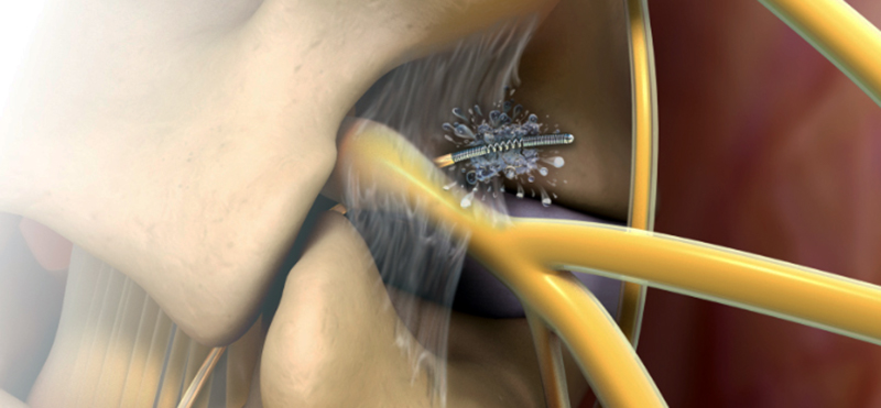
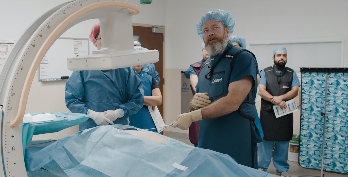
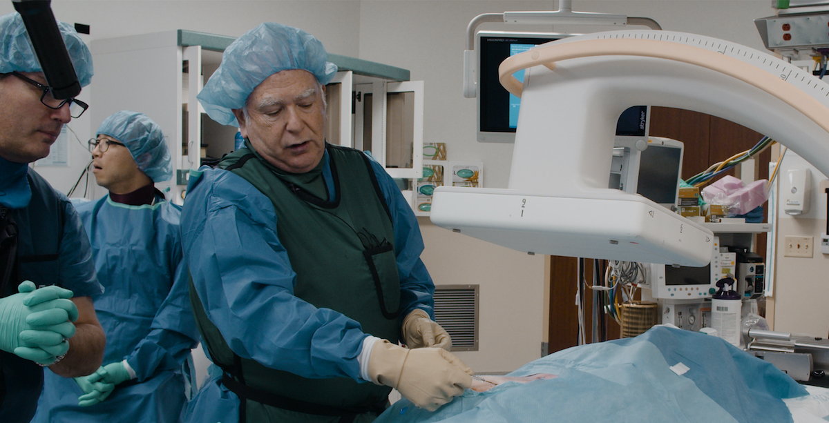
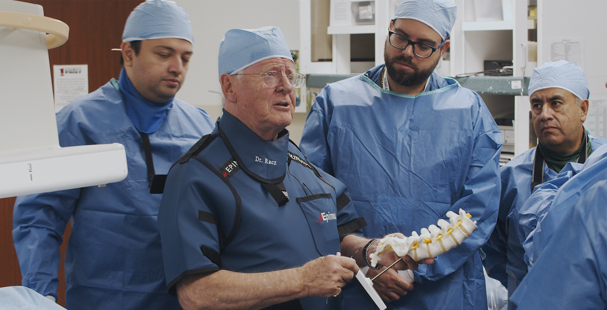
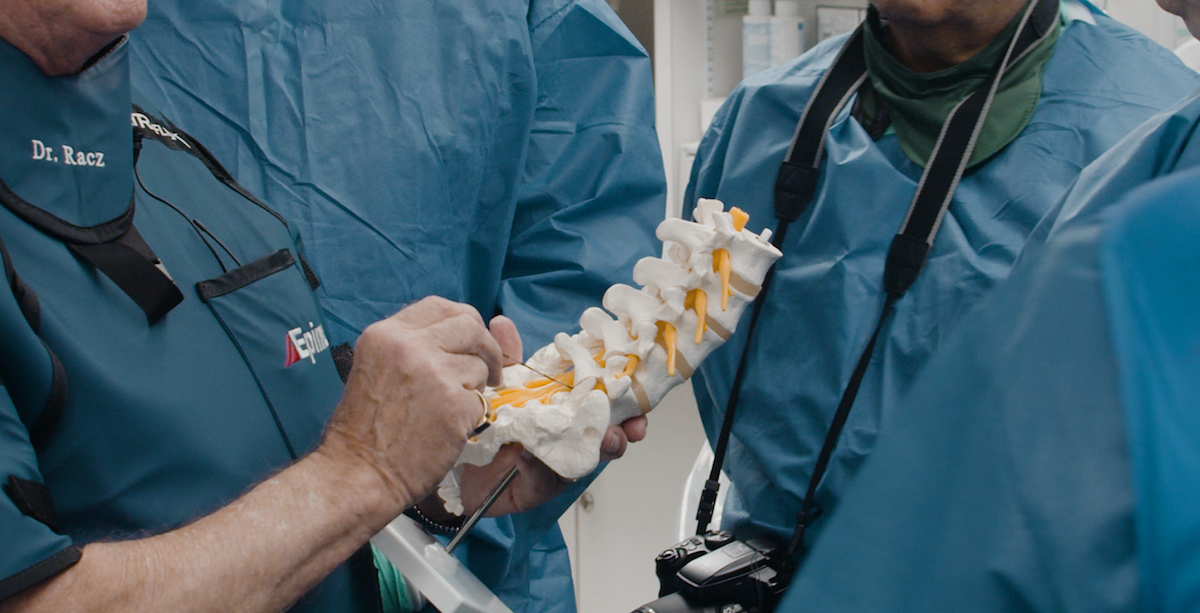
Similar to the topic as discussed in Pelvic Pain Procedures Scarring: Part Two, hydrodissection is carried out by introducing a 10cm, 20g Blunt Coude needle/cannula. The patient is in the prone position; the target for the needle placement is the tip of the ischial spine at the insertion of the iliosacral ligament. The needle tip slides posteriorly in the direction of the sacral nerve roots forming the pudendal nerve. The Injection of 5mL of contrast followed by 10mL of 150 units of Hylanex, 20 mg Triamcinolone and 8ml of even mixture of 0.25% Bupivicaine and 1% Lidocaine. The same volumes are injected anteriorly towards the inferior pudendal nerve distribution to free up the potentially scarred areas. If the pain relief is adequate but not long lasting, we proceed with pulsed radiofrequency of anterior and posterior direction of the pudendal nerve. The lesioning is 42 degree centigrade for 4-6 minutes in duration for each side using a 10cm, 10mm active tip curved Blunt Racz Finch Needle.
Interesting developments emphasize the delayed onset of scar formation in the presences of spreading abdominal wall hematoma. The hematoma also spread to the pelvic floor area leading to groin pain, pelvis with dyspareunia and weakness wasting of the legs. The groin pain has led to surgery of the ilioinguinal nerve that formed a painful neuroma. After the diagnostic blocks, cryolysis of the neuroma twelve years ago treated the condition and without the return of neuroma related pain. The delayed onset of scarring on the opposite left side became very obvious when 7 years later, the patient called to say that she has inability to move over the previous 8 months. During this time she had wasting of the leg from disuse and a painful knee. Surgical focus was addressing the knee pain with arthroscopy that resulted in bleeding in the knee and severe pain. Weakness and the evaluation of nerve function through EMG reported a silent vastus medialis muscle.
The patient had to travel from another state and it appeared very likely, because less bleeding occurred to the left side, the patient was undergoing the same scar formation of the femoral sheath in a delayed manner; additionally, she also had dyspareunia. We carried out hydro-dissection on the left side because the pelvic examination also confirmed the presence of a left sided painful pudendal nerve. There was no pain on the formerly painful right side.
The patient made a remarkable recovery of muscle strength of the femoral nerve innervated quadriceps muscles and during the 5 years, we did not have to repeat any procedures. The neurologist later revealed that he had interpreted the EMG report as an error and it was an artifact. Retrospectively, we believe the silent EMG reading was accurate, but just had not been formerly recognized therefore the reconsideration by neurologist calling it an artifact.